Tollund Man's head scanned in secrecy at Aarhus University
The world's best-preserved ancient human being has been scanned in an advanced scanner at the Department of Forensic Medicine. This has provided new information both about his internal condition and his cause of death.
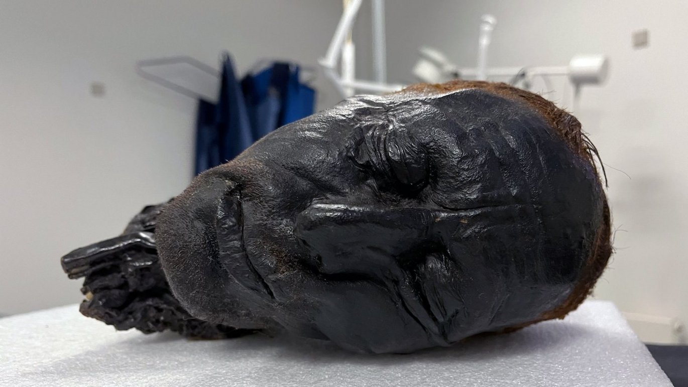
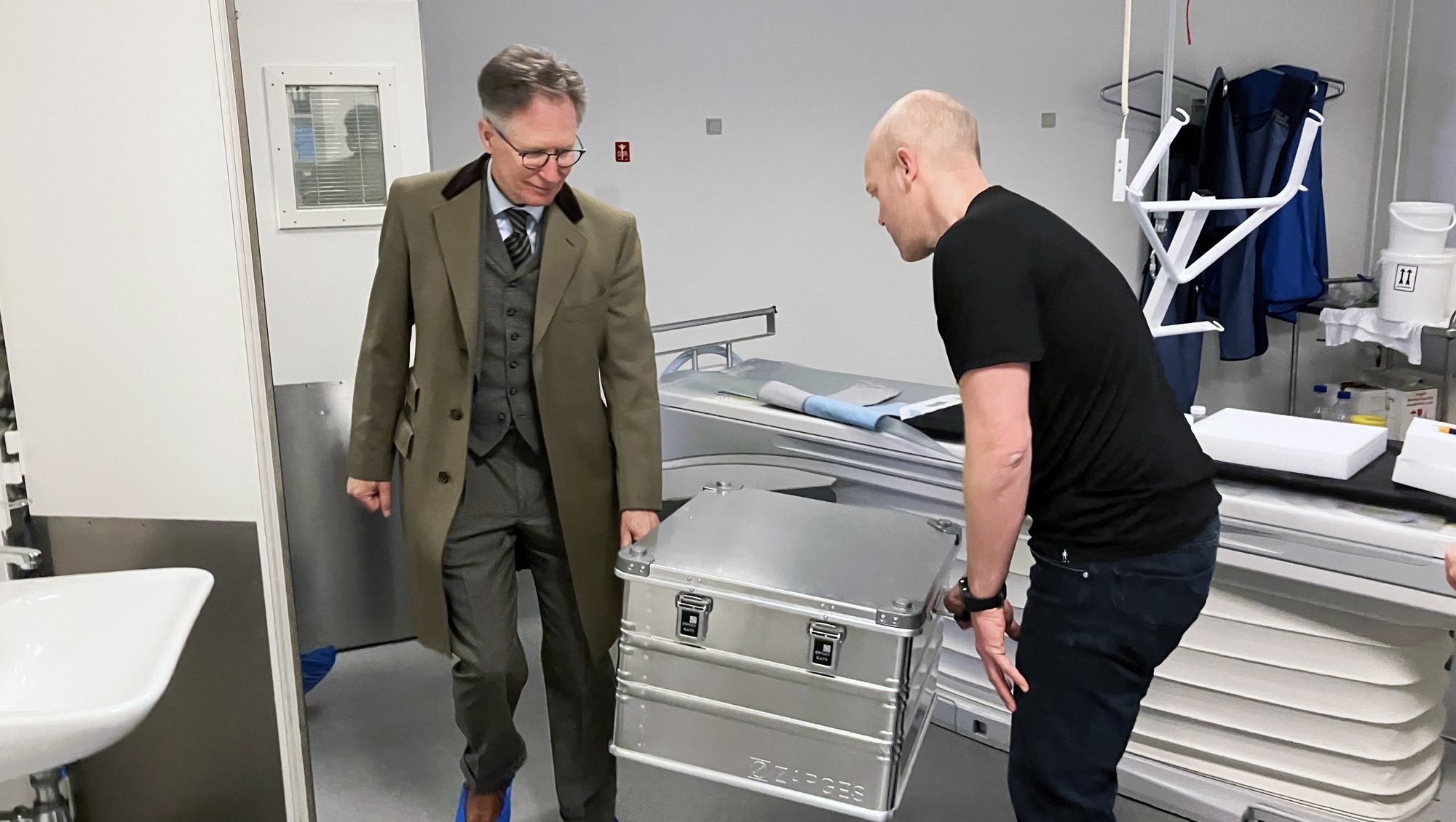
It is not every day that a severed human head is transported through Jutland in the trunk of a Volkswagen Golf Estate.
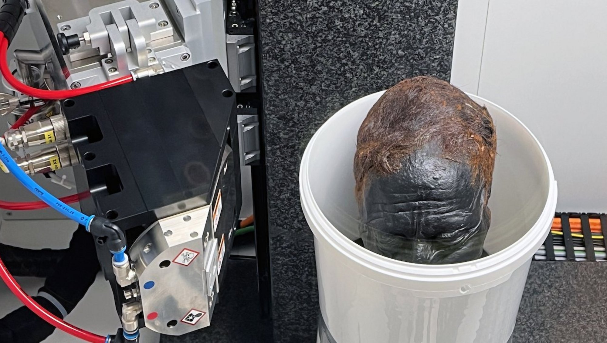
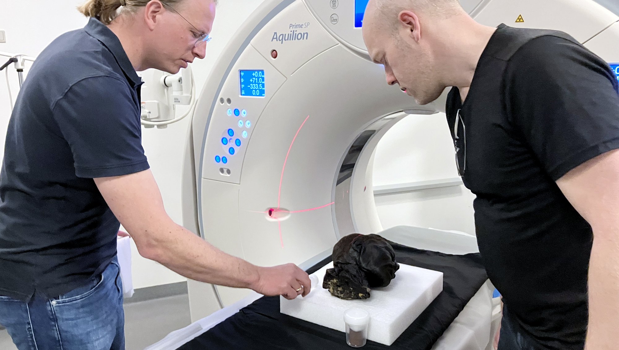
But on Monday 22 April, the head of the 2,400-year-old bog body Tollund Man was transported from Museum Silkeborg to the Department of Forensic Medicine at Aarhus University. Here, the famous head underwent thorough examinations in both a clinical CT scanner and in the university's new micro-CT scanner, which can produce highly detailed images.
Both researchers and people from the museum were awake for 24 hours while the scanning took place – but the result was worth it, says Ole Nielsen, director of Museum Silkeborg, where Tollund Man usually ‘lives’.
"We’re particularly curious about studying his teeth and neck. We suspect that he was hanged. This has been the main theory since Tollund Man was found with a leather rope around his neck in 1950. But previous studies in the form of X-rays and scans were not accurate enough to reject other theories – for example, that he was strangled," says Ole Nielsen.
About micro-CT scanning
As an employee at AU, are you interested in hearing more about the new micro-CT scanner - perhaps with a view to a collaboration?
Then contact Kasper Hansen or Henrik Lauridsen.
Aarhus University's new micro-CT scanner was purchased in an interdisciplinary collaboration between the Department of Forensic Medicine, the Department of Agroecology and the Department of Clinical Medicine.
The scanner was primarily financed by funds from the Carlsberg Foundation and the Department of Agroecology.
Micro-CT is a 3D imaging technique that uses X-rays to see inside an object, slice by slice. It takes a long time – in return, the images have an incredibly high resolution and a high level of detail.
The scanner is used to study a wide variety of materials, including clinical and forensic tissue samples, specimens from laboratory animals, soil samples and museum objects.
A completely intact eye
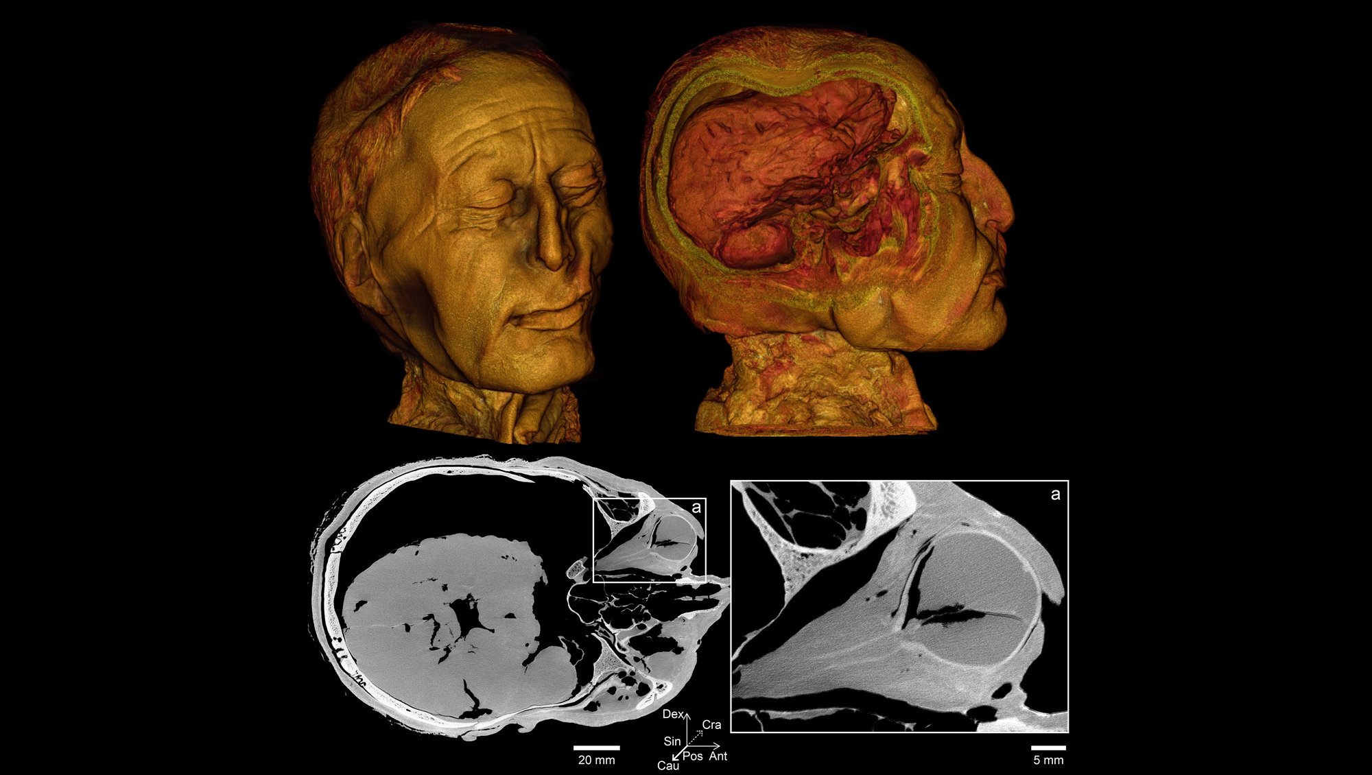
The museum director is thrilled with the detailed images of the Tollund Man's teeth and eyes, which cannot be seen from the outside because both his mouth and eyes are closed.
"One eye seems to be completely intact in terms of shape and optic nerves – that's amazing. The other eye is punctured," says Ole Nielsen.
"It's exceptional that his eyes and mouth aren’t completely damaged. Lips are one of the first things that are damaged when a corpse decomposes, but he is lying there with an almost Mona Lisa-like smile. We wouldn’t dream of forcing his mouth open to see his teeth, but now we can see them on the scan," he says.
Images of the Tollund Man's teeth can tell us if he’s had any dental diseases – and they can also help show how he lived his short life.
"If children experience long-term illness or hunger during childhood while their teeth are being formed, this will result in horizontal lines on their teeth. It can tell us whether he has had access to resources or whether there have been crises along the way. Does he have dental decay, does he have dental abscesses, and does he still have all his teeth? We want to know as much as possible about him," the museum director explains.
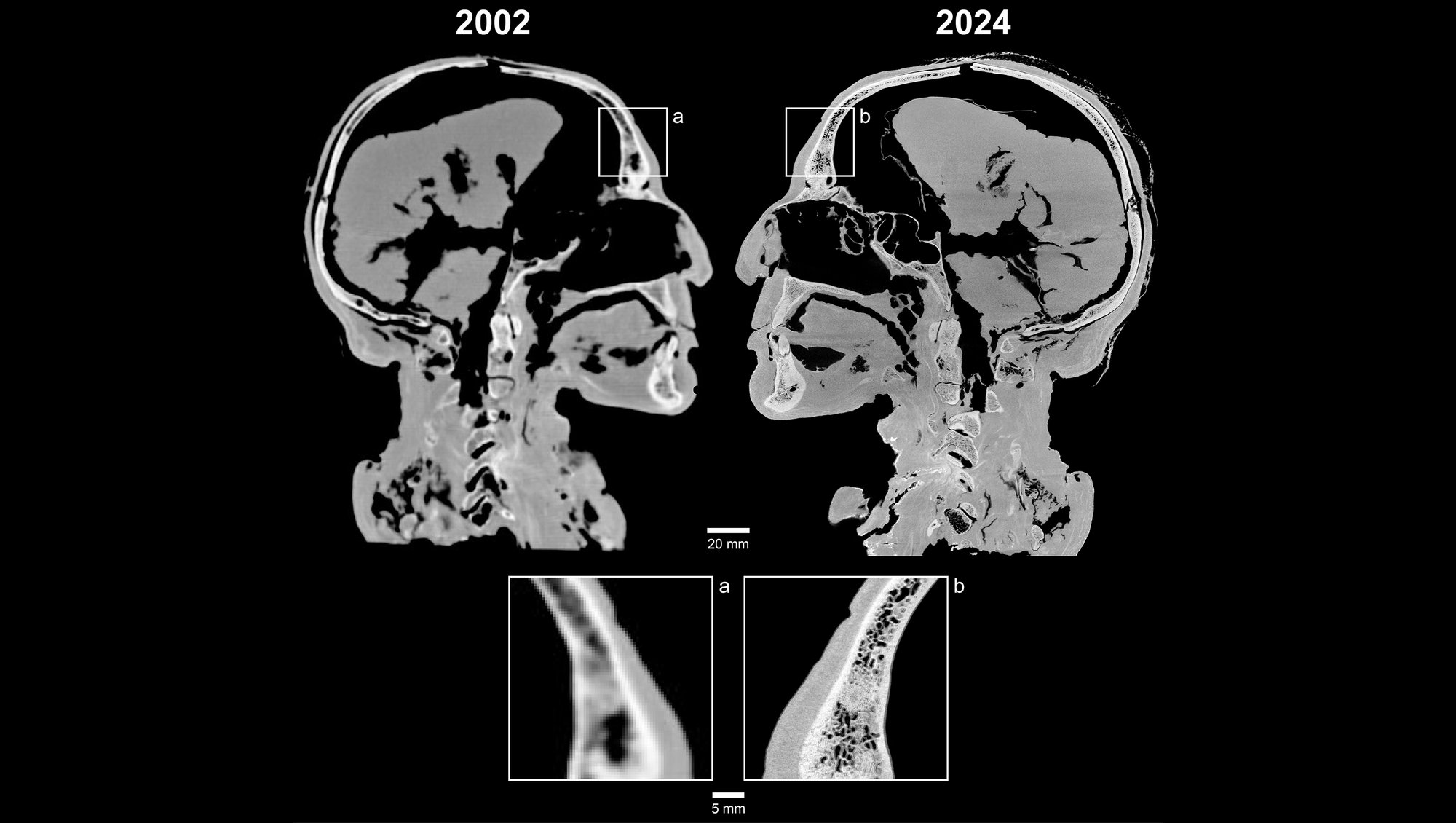
The head has been scanned once before – in 2002. On the old scans, the teeth appeared as vaguely outlined stumps. If you compare the new scans with the old ones, it’s just like getting a new pair of glasses, says Ole Nielsen. The resolution in the new images is 422 times higher than in the old ones. This makes a difference.
A police escort was used when the head was last scanned and transported from the glass showcase in the museum's main building where Tollund Man is on display for visitors from all over the world.
This time, the head was transported in a lined metal box in Museum Director Ole Nielsen's car. Conservator Lars Vig Jensen was also in the car, and the museum’s Head of Research Nina Helt Nielsen drove right behind them, ready to continue the transport of the head if anything should happen to the leading car.
The scan was carried out in secrecy.
"If there is too much fuss, it can break our concentration, which would make it more difficult to handle him correctly. Dropping him would be an irreparable loss. It was important that we had peace and quiet to work with the head in a proper and safe way," says the museum director.
About Tollund Man
Every year, thousands of people from all over the world travel to Museum Silkeborg to see the well-preserved body of the man who was found in a bog in 1950.
Tollund Man is approximately 2,400 years old. Over the years, he has been the subject of several scientific studies. Carbon-14 dating has shown that he died sometime between 405 and 380 BCE, i.e. in the early Iron Age. He was found with a leather rope around his neck.
His age is estimated to be 30-40 years, he was around 164-167 cm tall, and the size of his feet corresponds to a size 41. His hair was short and he was relatively freshly shaved when he was laid in the bog.
Tollund Man was probably in good health when he died. However, he suffered from both verrucas and intestinal worms.
Source: Museum Silkeborg
The results will be examined closely
Waiting to scan the Tollund Man's head with the brand new micro-CT scanner were Radiographer Christina Carøe Ejlskov Pedersen and Assistant Professor Kasper Hansen from the Department of Forensic Medicine, together with Assistant Professor Henrik Lauridsen from the Department of Clinical Medicine.
"The new scans are so impressive because of the good contrast, which makes it easy to see the difference between various tissues," explains Kasper Hansen.
"Because Tollund Man was lying in an acidic bog for more than 2,000 years, most of his bone minerals have been washed out. The contrast between bones and soft tissues is therefore less pronounced than normal. But with a micro-CT scanner, we can achieve better contrast on the different tissues and see a lot more details," the researcher explains and continues:
"The difference between an ordinary CT scanner and a micro-CT scanner is like switching from an old digital camera to a brand new one."
The new scanner differs from the university's other micro-CT systems because it can both scan small samples at ultra-high resolution and produce images of large objects – such as an entire head from an ancient human, Associate Professor Henrik Lauridsen points out.
"We use it to study everything from clinical and forensic tissue samples and specimens from laboratory animals to soil samples and museum objects," he says.
In the coming months, researchers from both Aarhus University and Museum Silkeborg will examine the results closely. And Tollund Man – he is now back at the museum, lying in his glass showcase and looking secretive.
Contact:
Assistant Professor Kasper Hansen
Aarhus University, Department of Forensic Medicine
Telephone: +45 87168304
Mail: kaha@forens.au.dk
Associate Professor Henrik Lauridsen
Aarhus University, Department of Clinical Medicine
Telephone: +45 61722106
Mail: henrik@clin.au.dk
Director Ole Nielsen
Museum Silkeborg
Telephone: +45 51214075
Mail: on@museumsilkeborg.dk
